Calm, dignified
experience

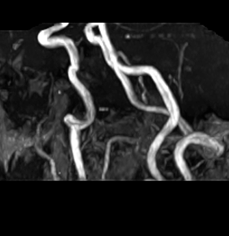
Carotid magnetic resonance angiography MRI scan
A carotid magnetic resonance angiography MRI scan can give a detailed view of the two carotid arteries on either side of the neck, the main blood vessels that deliver blood and oxygen to the head and brain.
Carotid MRA MRI scan
Medserena Carotid Magnetic Resonance Angiography MRI Scan
From £240.00
Magnetic Resonance Angiography scan of the Carotid; non-invasive procedure to help diagnose medical conditions relating to the arteries of the neck, price includes:
- Open and Upright MRI scan
- 20 minutes appointment
- Radiologist findings report
-
Images on USB at the end of the scanand available to NHS trusts via IEP on request
- Complimentary refreshments
Please wear metal free clothing and if possible, avoid wearing any jewellery. Alternatively, Medserena can provide you with a gown to change into for your scan. Scroll down for more Carotid
Artery Magnetic Resonance Angiography scan information.
many Scans Available within 48 hours
Superior healthcare service with every Private MRI scan
Little or no
waiting time
Largest MRI scan centres
Premium
refreshments
Watch TV while
scanning
Medical report included
About Carotid Magnetic Resonance Angiography MRI Scans
A carotid magnetic resonance angiography MRI scan can give a detailed view of the two carotid arteries on either side of the neck, the main blood vessels that deliver blood and oxygen to the head and brain. They supply the front part of the brain, which is the control centre for many important functions including speech, movement, and sensory feeling.
The main purpose of a carotid magnetic resonance MRA scan is to check for the build-up of fatty plaques or blood clots that can build up and cause blockages that put you at risk of a stroke or a mini stroke (known as a transient ischaemic attack (or TIA). MRI is an accurate imaging method for assessing the carotid arteries as it can measure vessel wall thickness.
Around 100,000 people a year in the UK suffer a stroke where the blood supply to the brain is blocked or reduced, and in up to one in four cases the cause is the narrowing of the carotid arteries. If the narrowing is discovered in time, surgery can be performed to correct the problem and prevent strokes and TIAs.
The advantage of an open carotid magnetic resonance angiography MRI scan is that you can sit in a comfortable upright scanner with equipment around your neck. The scan takes 15 minutes to complete and examines the blood vessels from the base of the brain to the top of the aorta, the main artery in the body. Having an open scan may be particularly reassuring for you if you suffer from fear or anxiety associated with confined spaces (claustrophobia), particularly tunnel MRI scans.
What conditions can a carotid magnetic resonance angiography MRI scan detect?
- Carotid artery disease: This is where the carotid arteries narrow, called carotid stenosis due to the build-up of fatty deposits causing plaques to build up. Risk factors for developing carotid artery disease include older age, high blood pressure, diabetes, raised cholesterol, smoking and heart disease. These plaques can narrow the arteries and if they break away, they can cause a clot to block the artery. If the clot then breaks away from the plaque it becomes a pulmonary embolism. These clots (thrombus) can potentially travel to the brain causing a stroke. If narrowing is detected it can be tackled with an operation called a carotid endarterectomy to remove the fatty plaque deposits.
- Carotid dissection/tears: The inside layers of the carotid artery can tear, letting blood pool, causing the artery wall to bulge, pressing on surrounding nerves or tissue, or causing a blood clot to form. Symptoms can include headaches, neck, eye or scalp pain, droopy eyelids, and a pulsing sound in the ear. It can be caused by an accident such as a car crash or have no apparent cause.
- Predicting the risk of future heart attacks and strokes: A study published in the medical journal Radiology found that MRI imaging of the carotid arteries can predict vulnerability to future heart attacks and strokes.
Other benefits of a Medserena carotid magnetic resonance angiography scan
Open MRI scanners are a stress-free alternative to using a conventional enclosed tunnel MRI scanner, providing comfort and reassurance for people who suffer from anxiety or claustrophobia. Sitting upright is more comfortable for patients and the open front means patients can speak to a friend or relative or watch television throughout as distraction.
Open MRI scans can also accommodate larger/ heavier patients who might have difficulty fitting comfortably into a conventional tunnel scanner, as they can take weights of up to 35 stone (226kg). However, suitability will depend on the patient’s build and the area of anatomy that needs to be scanned.
- To book a Medserena carotid magnetic resonance angiography MRI scan direct in London or Manchester, go to www.medserena.co.uk
FAQs
The Upright MRI is truly open. There are no tunnels, no narrow tubes. The system is particularly quiet, the examination is comfortable and does not trigger feelings of being in a confined space. This means that the Upright MRI is particularly tolerated by patients who suffer from “claustrophobia”.
Because the system offers you an unrestricted view, you can watch TV or see DVD movies on a large screen during the scan. Wearing headphones – as with other MRI systems – is usually not necessary.
According to the current state of knowledge, there is no danger to the patient’s health as magnetic resonance imaging only uses magnetic fields and radio waves.
Metallic foreign bodies within the patient, such as fixed dental prosthesis, artificial joints or metal plates after treatment for a fracture do not usually pose any danger. However, it is important to clarify that the implants you use are MRI-compatible before the examination.
MRI (Magnetic Resonance Imaging) utilises a large magnet, radio waves and a computer to form images of your body. It is non-invasive, painless and does not use any ionising radiation.
Our truly open MRI can scan you in different positions. Through the utilisation of a specially designed MRI system we can offer weight-bearing scans – sitting or standing. The design of the system allows the patient to be positioned in different postures (e.g. flexion or extension) so that the patient may be examined in the position where they experience pain. The reason to do this is that some pathologies are underestimated or even not seen in a conventional supine MRI scan. The technique has value in many applications: e.g. spine, knees, hips, ankles. This has been proven in scientific studies and documented in peer reviewed publications.
In addition, it offers the possibility of performing an MRI scan on patients who could not otherwise tolerate the examination. This may include the claustrophobic patient, who benefits from the truly open nature of the equipment, and the severely kyphotic patient or emphysema sufferer who simply cannot lie down. It can also facilitate scanning of large patients who struggle to fit conventional ‘bore’ MRI scanners.
Of course, we have a comfortable waiting area but if you want them to stay in the scan room with you, they will also need to fill out a safety questionnaire. There is enough space for a companion. The person can even hold your hand and communicate with you during the examination. This is particularly beneficial when examining teenager.
This depends above all on which part of the body needs to be examined. In the Upright MRI, special examinations can be carried out in various body positions. The entire scan generally takes between 30 and 45 minutes. However, since you have the opportunity to watch TV or DVD, this time will go by much quicker.
Eat and drink normally and, unless your doctor tells you otherwise, please continue taking medications as normal. If you have any special needs (e.g. wheelchair access) please inform us when making the appointment.
Your appointment confirmation; referral letter/form; Medical Insurance details if applicable. We accept all major debit/credit cards.
We will provide a gown/clothing for you to wear when you are scanned. If you prefer to wear your own, please ensure that you wear or bring clothing without any metal fasteners, zips or under-wiring as these cannot be worn in the scan room. The changing room can be locked for safe storage of your possessions.
You will be able to walk into the scanner. It has no tunnel or bore. You will be able to hear us and talk with us during your scan if necessary-and we will be able to see you at all times. Due to its open nature, you will even be able to watch TV or a DVD whilst having the scan. Depending on which part of you is being scanned, you may be asked to sit or stand, and assume different postures (for example bending forward.) The radiographer may place a receiver “coil” around the relevant area of your body. You will need to remain very still while the acquisition is done in order to prevent blurring of the images. You will hear some tapping from the scanner but in general it is much quieter than many other MRI scanners.
You will not feel anything while having the scan. There is no pain or unusual feeling of any type and you will experience no after effects.
YES. There are some things that can prevent you from having an MRI scan. You will be asked to complete a safety questionnaire on arrival at the Centre which will cover the contra-indications-but if you are making an appointment and any of the factors below affect you, please discuss this with us in advance as it may save you a wasted trip.
Contra-indications can include:
- Pacemaker
- IUDs
- Surgical clips
- Pregnancy
- Metal fragments in the body
- Metal pins/plates/screws
- Joint replacements
- Metal objects in eyes
- Cochlear implants
- IVC filters
- Metal heart valves
- Penile implants
It is also important to tell us if you have any tattoos or piercings.
Watches, jewellery, coins, keys, cigarette lighters, penknives, credit cards. piercings, hairgrips, wigs, nicotine patches, and hearing aids must be removed.
Your scan will be reported by a Consultant Radiologist. It will normally be available in a couple of days unless needed urgently. The images and report will be sent to your referring practitioner. If you have a follow up appointment, please make us aware of the details so we can ensure the report and images are available in time.